Introduction to Animal Cell Parts: Animal Cell Parts Coloring Page

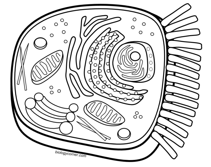
Animal cell parts coloring page – Animal cells are the fundamental building blocks of animals, exhibiting a complex internal structure responsible for carrying out various life processes. Unlike plant cells, they lack a rigid cell wall and chloroplasts, but possess a variety of organelles that work together to maintain cellular function and contribute to the overall health of the organism. Understanding the structure and function of these organelles is crucial for comprehending the complexities of animal life.Animal cells are characterized by their dynamic and flexible nature, allowing for a range of movements and interactions.
This fluidity is supported by the cytoskeleton, a network of protein filaments that provides structural support and facilitates intracellular transport. The organelles within are not static; they constantly move and interact, reflecting the cell’s active metabolism and responsiveness to its environment.
Animal Cell Organelles and Their Functions
The major organelles within an animal cell each play a specific role in maintaining cellular life. The nucleus, for instance, houses the cell’s genetic material (DNA), controlling gene expression and directing cellular activities. The mitochondria, often called the “powerhouses” of the cell, generate energy in the form of ATP through cellular respiration. The endoplasmic reticulum (ER), a network of membranes, synthesizes proteins and lipids.
Yo, so I was totally vibing with this animal cell parts coloring page, like, seriously detailed. Then I remembered I also gotta check out some chainsaw man anime coloring pages for a total change of pace, ’cause Denji’s chainsaw is way more hype than a nucleus. But yeah, back to the animal cell – gotta ace that biology homework, right?
The Golgi apparatus processes and packages proteins for secretion or transport within the cell. Lysosomes break down waste materials and cellular debris. The ribosomes, responsible for protein synthesis, can be found free-floating in the cytoplasm or attached to the ER. Finally, the cell membrane encloses the entire cell, regulating the passage of substances in and out.
Animal Cell Parts Commonly Featured in Coloring Pages
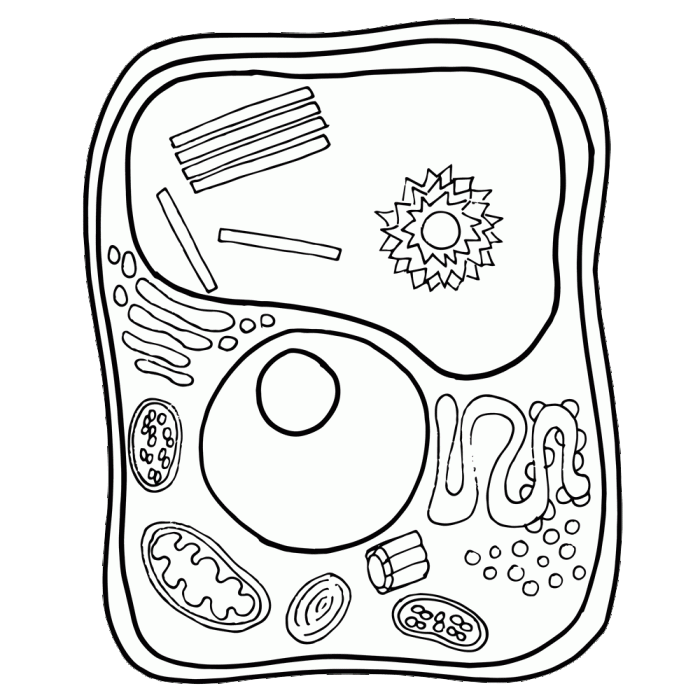
Typically, coloring pages depicting animal cells focus on the major organelles readily visible under a microscope. A simplified representation is often used for educational purposes.This usually includes a depiction of the cell membrane, outlining the cell’s boundary. The nucleus, typically shown as a large, centrally located structure, is another key component. Mitochondria are often represented as small, bean-shaped structures scattered throughout the cytoplasm.
The endoplasmic reticulum might be depicted as a network of interconnected tubules and sacs, while the Golgi apparatus may be simplified as a stack of flattened sacs. Ribosomes are often omitted due to their small size and difficulty in visualization. Lysosomes may or may not be included, depending on the complexity of the coloring page. The cytoplasm, the jelly-like substance filling the cell, serves as the background for these organelles.
In some more detailed coloring pages, the centrosome, involved in cell division, might also be included.
Organelle Descriptions for the Coloring Page

This section provides detailed descriptions of the key organelles found within an animal cell. Understanding their individual functions is crucial to grasping the overall complexity and efficiency of cellular processes. Each organelle plays a specific and vital role in maintaining the cell’s life and function.
The Nucleus: Control Center of the Cell, Animal cell parts coloring page
The nucleus is the cell’s control center, housing the genetic material – DNA – organized into chromosomes. DNA contains the instructions for building and maintaining the entire organism. The nucleus is enclosed by a double membrane called the nuclear envelope, which regulates the passage of molecules in and out. Within the nucleus, a dense region called the nucleolus is involved in ribosome synthesis.
During cell division, the DNA replicates and the chromosomes condense, ensuring each daughter cell receives a complete set of genetic instructions. This precise replication and distribution are fundamental to growth and reproduction.
Mitochondria: Powerhouses of the Cell
Mitochondria are often referred to as the “powerhouses” of the cell because they are responsible for cellular respiration. This process converts the chemical energy stored in glucose into a usable form of energy called ATP (adenosine triphosphate). ATP fuels various cellular activities, from muscle contraction to protein synthesis. Mitochondria have a double membrane structure, with the inner membrane folded into cristae, increasing the surface area for ATP production.
The number of mitochondria in a cell varies depending on the cell’s energy demands; cells with high energy requirements, such as muscle cells, have many more mitochondria than less active cells.
Endoplasmic Reticulum and Golgi Apparatus: Protein Synthesis and Transport
The endoplasmic reticulum (ER) and Golgi apparatus work together in protein synthesis and transport. The rough ER, studded with ribosomes, is the site of protein synthesis. Ribosomes translate the genetic code from mRNA into polypeptide chains, which fold into functional proteins. These proteins then move to the smooth ER, which is involved in lipid synthesis and detoxification. From the ER, proteins are transported to the Golgi apparatus, a stack of flattened sacs.
The Golgi apparatus modifies, sorts, and packages proteins into vesicles for transport to their final destinations, either within the cell or outside the cell via exocytosis.
Lysosomes: Waste Disposal System
Lysosomes are membrane-bound organelles containing digestive enzymes. They act as the cell’s waste disposal system, breaking down waste products, cellular debris, and foreign substances such as bacteria. These enzymes work best in acidic environments, which are maintained within the lysosomes. Lysosomes play a crucial role in recycling cellular components, ensuring the cell maintains a healthy and efficient internal environment.
Malfunctions in lysosomes can lead to various diseases due to the accumulation of undigested materials.
Cell Membrane: Maintaining Cell Integrity
The cell membrane, also known as the plasma membrane, is a selectively permeable barrier that encloses the cell’s contents. It regulates the passage of substances into and out of the cell, maintaining the cell’s internal environment. The membrane is composed of a phospholipid bilayer, with embedded proteins that perform various functions, including transport, cell signaling, and cell adhesion.
The selective permeability of the membrane is crucial for maintaining homeostasis, ensuring the cell has the necessary nutrients and expels waste products effectively. The integrity of the cell membrane is essential for the survival of the cell.
Illustrative Examples
To further solidify understanding of animal cell components, let’s visualize key organelles through detailed descriptions. These descriptions will help you accurately represent these structures in your coloring page.The following sections provide detailed descriptions of illustrative examples of a nucleus, mitochondrion, and endoplasmic reticulum. These descriptions aim to provide a comprehensive understanding of their structures and functions, enhancing the accuracy of your artistic representation.
Nucleus
Imagine a large, roughly spherical structure, the nucleus, occupying a central position within the cell. Its defining feature is the nuclear envelope, a double membrane studded with nuclear pores. These pores are crucial for regulating the transport of molecules between the nucleus and the cytoplasm. Within the nucleus, a prominent, darker-staining region, the nucleolus, is visible. This is the site of ribosome biogenesis.
The remaining space is filled with a complex network of DNA and associated proteins, collectively known as chromatin. In a detailed image, the chromatin would appear as a diffuse, thread-like material, which condenses into distinct chromosomes during cell division. The nuclear membrane’s double layer should be clearly depicted, with the nuclear pores appearing as small openings puncturing the membrane.
Mitochondrion
Visualize an oval-shaped organelle, the mitochondrion, often described as the “powerhouse” of the cell. It is characterized by a double membrane system: an outer membrane and an inner membrane. The inner membrane is extensively folded into numerous cristae, which greatly increase the surface area available for cellular respiration. These cristae appear as shelf-like or finger-like projections extending into the mitochondrial matrix, the space enclosed by the inner membrane.
A detailed image should clearly distinguish the outer membrane from the inner membrane, showcasing the numerous cristae and the relatively smooth outer membrane. The matrix itself should be represented as a space filled with enzymes and other molecules involved in energy production.
Endoplasmic Reticulum
Envision an extensive network of interconnected membranous sacs and tubules extending throughout the cytoplasm – this is the endoplasmic reticulum (ER). There are two distinct types: rough ER and smooth ER. The rough ER appears rough due to the presence of ribosomes attached to its surface. These ribosomes are responsible for protein synthesis. In contrast, the smooth ER lacks ribosomes and appears smoother.
It is involved in lipid synthesis, detoxification, and calcium storage. A detailed image would illustrate the difference in appearance between the rough and smooth ER, showing the ribosomes studded on the surface of the rough ER and the smooth, tubular structure of the smooth ER. The interconnectedness of the ER network should also be emphasized, demonstrating its extensive reach within the cell.
